|
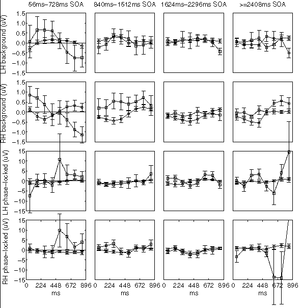
For the SSVEP data, analysis of variance revealed an effect of SOA × group (F(3, 54) = 3.06, p = 0.0359). Group effects on background amplitudes were more pronounced than those on the SSVEP itself, owing to less variability in the data: for the background amplitudes there were strong effects of latency × group (F(7, 126) = 6.38, p < 0.0001) and of SOA × latency × group (F(21, 378) = 3.57, p < 0.0001). The complete data set is expressed graphically in figure 1; for cells that contribute heavily to these statistical effects, means and standard errors are given in the text below. Since the quantity represented is the difference in amplitude between left targets (which cue rightward shifts) and right targets (which cue leftward shifts), positive values indicate greater response to left targets and negative values indicate greater response to right targets.
|
At short SOAs, people with autism showed much greater SSVEP response to left targets (rightward shifts) than to right targets (leftward shifts), beginning at about 500ms post-target (figure 1, column 1, bottom two panels). This heightened response occurred in both hemispheres, but was more consistent in the right hemisphere: for the 56ms-728ms SOA range, at 600ms post-target, right-hemisphere mean difference amplitude was 6.8±6.5µV for people with autism versus -1.4±0.8µV for controls, while the left-hemisphere means were 3.3±4.9µV for autism and 0.1±0.4µV for controls. (All ranges are standard error of the mean.) In contrast to this large, bihemispheric effect in autism, the normal pattern of SSVEP modulation consisted of a small left-right difference that increased (i.e., became greater for rightward shifts) in the left hemisphere but decreased (i.e., became greater for leftward shifts) in the right hemisphere. People with autism did not manifest such separate and opposite effects in the two separate hemispheres; instead, the hemispheres behaved similarly to each other, each responding more highly during rightward shifts than during leftward shifts.
SSVEP differences at long SOAs (lower right quadrant of figure 1) were highly variable in the autistic subjects, oscillating between positive and negative signs. For SOA ≥ 2408ms, right-hemisphere mean difference amplitude at 600ms was -14.0±13.2µV for autism versus 2.4±1.1µV for controls. The corresponding values for the left hemisphere were -6.1±5.7µV and 0.1±0.5µV. These large and disordered amplitudes in the case of autism were utterly dissimilar to the normal pattern in which well defined and oppositely directed modulations in the two hemispheres are evident as early as 300ms post-target.
In the background (non-phase-locked) EEG, at short SOAs people with autism showed strongly decreasing response to left targets (rightward shifts) and increasing response to right targets (leftward shifts) (figure 1, column 1, top two panels). Again the direction of the effect was the same in both hemispheres. The control subjects, in contrast, manifested a weak, decreasing left-right difference in the left hemisphere but a stronger, increasing trend in the right hemisphere. For the 56ms-728ms SOA range, by 700ms post-target the mean left-right difference had fallen to -0.89±0.37µV in autism as compared to 0.32±0.26µV in controls, in the right hemisphere; and to -0.75±0.65µV for autism as compared to 0.03±0.07µV for controls, in the left hemisphere.
At longer SOAs, this pattern of bihemispheric decrease in difference amplitude of the background EEG disappeared and, in the case of the right hemisphere, amplitude became high in response to left targets (rightward shifts) throughout the epoch (column 2, panel 2). For the 840ms-1512ms SOA range, by 300ms post-target the amplitude in controls was -0.44±0.17µV whereas in autism it had increased to 0.54±0.40µV. In addition, right-hemisphere background amplitude modulation that was quite pronounced in control subjects in the two longest SOA ranges was utterly absent in autism (columns 3 and 4, panel 2). For SOA ≥ 2408ms, difference amplitudes in controls climbed from -0.55±0.22µV at 400ms post-target to 0.56±0.26µV at 700ms post-target, whereas for people with autism the amplitudes remained flat, from 0.09±0.22µV at 400ms to 0.0±0.08µV at 700ms.
|
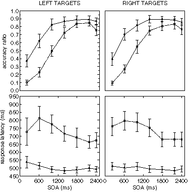
The behavioural data are displayed in figure 2. As expected, there were highly significant effects of SOA on accuracy (F(6, 66) = 43.27, p < 0.0001) and on response latency (F(6, 66) = 3.49, p = 0.0047). The ratio of correct target detections to total number of detection opportunities was lower for the autistic group than for the normal group (0.58±0.04 for autism and 0.77±0.06 for controls, F(1, 18) = 6.48, p = 0.0203), and attained its maximum at a longer SOA (SOA × group effect, F(6, 108) = 9.53, p < 0.0001). The response latency also was greater for the autistic group (728±43ms for autism and 500±20ms for controls, F(1, 18) = 24.21, p = 0.0001), and decreased more rapidly with increases in SOA (SOA × group effect, F(6, 108) = 4.01, p = 0.0012). Interestingly, the response latency was somewhat lower for correctly detected targets in the shortest SOA range than for the next shortest.