fMRI results: normal subjects, extrastriate
Again, since this is a region-of-interest analysis within individuals, I don't have any flashy coloured parametric maps on averaged brains to show you. However, I think that the numbers by themselves tell a more compelling story.

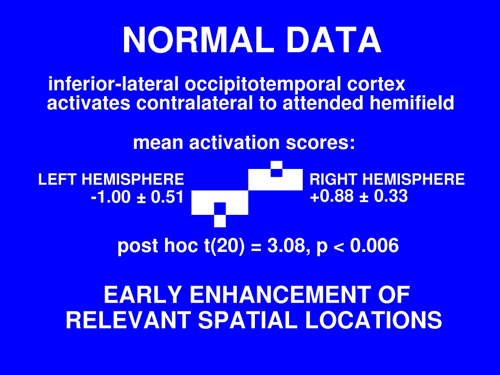
First, the normal data. Results for the occipitotemporal region agree very well with what we found in the EEG study: each hemisphere is more active when attention is directed within the contralateral visual hemifield.

Our ideal waveform is 1 when subjects are attending left and 0 when subjects are attending right. So a correlation with left attention produces a positive activation score, and a correlation with right attention produces a negative activation score.


[previous] [contents] [next]
`fMRI Evidence for Generalised Arousal as a Substitute for Early Selection in Autism during Conditions of Shifting Visual Spatial Attention', Matthew Belmonte, 10 November 2001