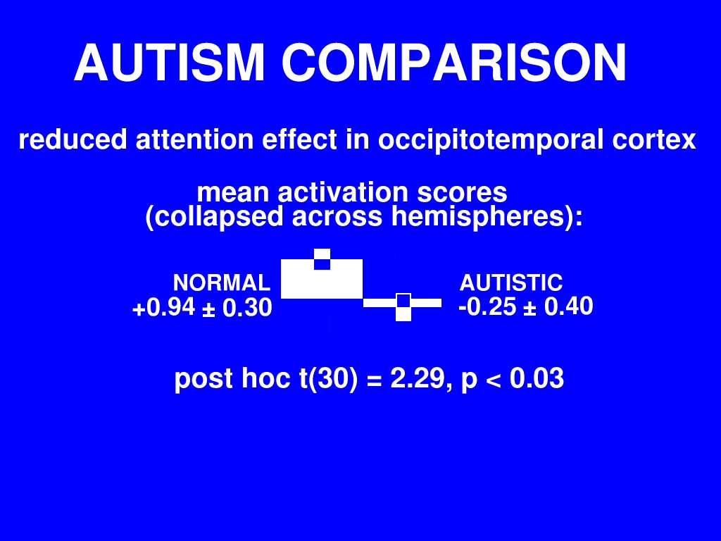
In this figure we've collapsed the data across hemisphere by multiplying the left-hemisphere measures by negative one and adding them to the right-hemisphere measures.
Whereas normal subjects show a strong, selective activation contralateral to the attended hemifield, in autism this selective activation is absent.
I want to mention again here that although there was a gender-associated trend within the normal sample, that trend cannot account for this difference since within the normals we found that males showed a greater degree of lateralised activation, and in our autism sample, which is weighted towards males, we see an absence of lateralised activation.